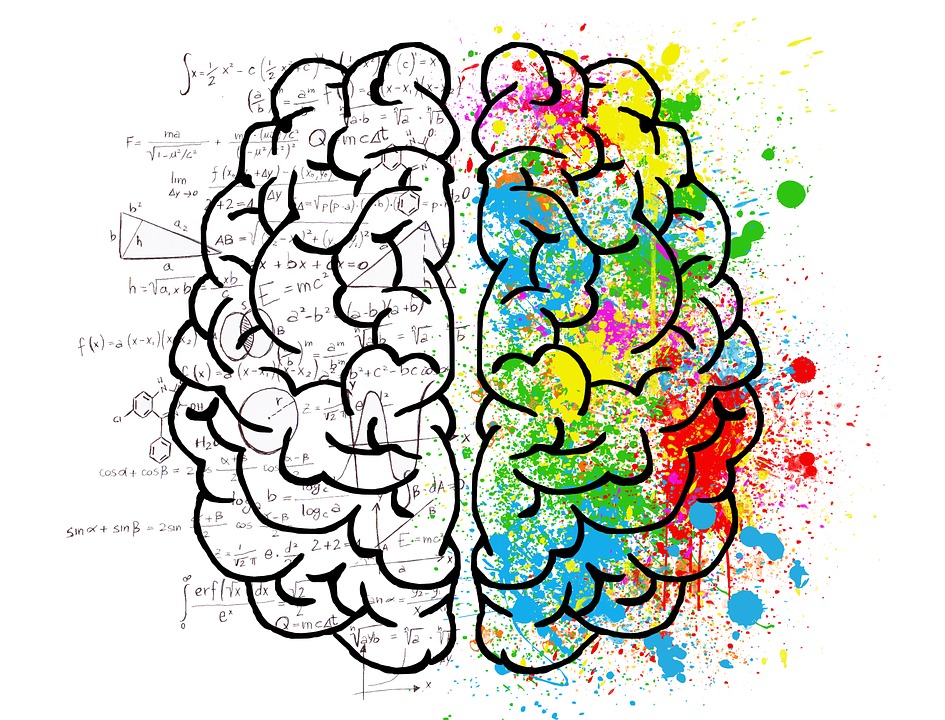
‘Split brain patients’ are individuals who have had the major connection between the two hemispheres of their brain severed, usually in an attempt to lessen the frequency of fitting in extreme cases of epilepsy. Since the 1940s, it has been accepted that the cutting of one’s corpus callosum (the connection between the two hemispheres of the brain) is likely to produce both desirable and undesirable effects. However recent research into this has suggested that there might be more to this peculiar practice than was first thought.
The treatment of epilepsy can effectively act as a guideline as to the development of psychological methods of studying the brain, particularly highlighted by the recent discovery that the surgical process of cutting the connection between the hemispheres does not create two separate, conscious structures within the brain, but instead prevents certain functions as the two hemispheres are no longer able to communicate. This process originally aimed to prevent the storm of fit-inducing electricity in the patient’s brain from passing from one hemisphere to the next. In many cases, the treatment was successful. But, as is often the case in the world of science, the most interesting result was not its intended effects, but the unintended ones – and what they’ve taught us about the localisation of function within the human brain.
Our understanding on this topic began with research conducted by Sperry and Gazzaniga, who studied the relatively few patients who underwent the procedure as a last resort. Their (anonymous) first patient had been experiencing seizures since being hit in the head by the butt of a German soldier’s rifle in WW2. After the separation procedure, an experiment was set up in which an image was flashed either in his left or right visual field. He had a button for each hand to press when he saw an image on the corresponding side. Everything appeared normal apart from, during instances in which an image flashed in his left visual field, he said he saw nothing but his left hand kept pressing the button. This is because each hemisphere of the brain controls the opposite side of the body. Therefore when an image appeared in the patient’s left visual field, the information was processed by the right hemisphere which, unlike the left hemisphere, does not contain a language processing centre and so couldn’t verbalise what he had seen, despite his brain having registered the presence of the image.
Each hemisphere of the brain controls the opposite side of the body.
Another particularly notable case study of theirs was that of a young boy asked about his favourite girlfriend at school. When the question was presented to him visually in his left visual field, the boy shrugged to indicate that there was no question, but laughed and used his left hand to spell LIZ slowly in scrabble pieces, indicating that his right hemisphere had recognised the question presented to him, yet the verbal processing centres in his left hemisphere remained unaware.
If anything can be taken from this, it’s that we should all be grateful for the brain scanning techniques and medication that have replaced such practices, allowing us to freely experience and verbalise our experiences, all whilst avoiding invasive and potentially unnecessary brain surgery.
By Abby Drew
image source: https://www.scienceatcal.berkeley.edu

